Lanthanum »
PDB 1djg-9ceq »
2i18 »
Lanthanum in PDB 2i18: The Refined Structure of C-Terminal Domain of An Ef-Hand Calcium Binding Protein From Entamoeba Histolytica
Lanthanum Binding Sites:
The binding sites of Lanthanum atom in the The Refined Structure of C-Terminal Domain of An Ef-Hand Calcium Binding Protein From Entamoeba Histolytica
(pdb code 2i18). This binding sites where shown within
5.0 Angstroms radius around Lanthanum atom.
In total only one binding site of Lanthanum was determined in the The Refined Structure of C-Terminal Domain of An Ef-Hand Calcium Binding Protein From Entamoeba Histolytica, PDB code: 2i18:
In total only one binding site of Lanthanum was determined in the The Refined Structure of C-Terminal Domain of An Ef-Hand Calcium Binding Protein From Entamoeba Histolytica, PDB code: 2i18:
Lanthanum binding site 1 out of 1 in 2i18
Go back to
Lanthanum binding site 1 out
of 1 in the The Refined Structure of C-Terminal Domain of An Ef-Hand Calcium Binding Protein From Entamoeba Histolytica
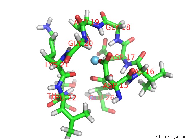
Mono view
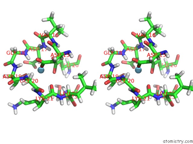
Stereo pair view
Mono view
Stereo pair view
A full contact list of Lanthanum with other atoms in the La binding
site number 1 of The Refined Structure of C-Terminal Domain of An Ef-Hand Calcium Binding Protein From Entamoeba Histolytica within 5.0Å range:
|
Reference:
S.M.Mustafi,
S.Mukherjee,
K.V.Chary,
G.Cavallaro.
Structural Basis For the Observed Differential Magnetic Anisotropic Tensorial Values in Calcium Binding Proteins. Proteins V. 65 656 2006.
ISSN: ISSN 0887-3585
PubMed: 16981203
DOI: 10.1002/PROT.21121
Page generated: Tue Aug 13 01:47:48 2024
ISSN: ISSN 0887-3585
PubMed: 16981203
DOI: 10.1002/PROT.21121
Last articles
Cl in 3MSWCl in 3MS9
Cl in 3MSU
Cl in 3MSJ
Cl in 3MSL
Cl in 3MSK
Cl in 3MN1
Cl in 3MS7
Cl in 3MR1
Cl in 3MQE